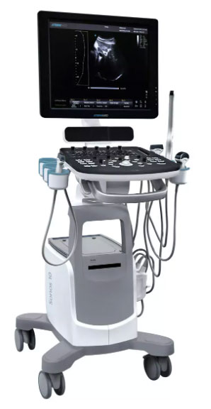
Ultrazvuk Sonos 10

Karakteristike
Full display Mode Full-screen mode bez gubitka rezolucije slike, pruža Vam više detalja za što precizniju diijagnostiku. HIP Graf koristi graf za dijagnostiku kukova, pomažući doktoru za davanje lakše i tačnije dijagnoze tokom skrininga dečijih kukova. Različit ugao indicates different level of hip deformity, which is easier and more obvious to see with the aid of the graph. (I, II, D llla, lllb). Auto IMT Function Automatically traces the intima and measures the thickness of the intima. This allows you to measure the intima faster, more easily and more accurately. Super Needle With Super Needle, clinicians can see needle inside tissue more clearly during medical procedures. (Needle angle up to ±30°)
Advanced Technologies
X-contrast
- The Sonos 10 allows one-touch user-adjusted contrast resolution based upon differences in tissue density.
- Enhance, Normal, and Suppress settings increase or decrease contrast resolution, based on the tissue type and user preference.
FHI
- FHI is an innovative harmonic imaging technology that uses multiple transmission and receiving methods based on the patients’ size and weight. This allows the Sonos 10 to maintain image resolution when imaging larger patients.
- Traditional Tissue Harmonics and Phased Harmonics compromise image quality and resolution when penetration is increased.
- FHI technology greatly improves diagnostic abilities and clinical confidence in larger, difficult-to-image patients.
Q-Beam
- Compared to the traditional dual-beam former on most ultrasound machines, the Sonos 10 uses quad-beam for ultrasound
- Doubles the volume of signals received over traditional methods, increasing image resolution and generating more accurate images.
- Produces higher frame rates, ensuring better diagnostic confidence and efficiency, especially for moving organs.
Q-Flow
- This adaptive color detection technology can automatically adjust the assessment of color signal and noise according to different tissues.
- As a result, color sensitivity of low-velocity flow is significantly enhanced.
Specifications
- High resolution 19” LED display
- Reporting system
- Intelligent Zoom
- Pulse Wave Doppler
- Image review system
- 3 active probe ports (standard)
- Color Doppler Flow Imaging
- Power Doppler Flow Imaging
- Multi-language screen display
- Automatic Image Optimization
- patient information management system
- Directional Power Doppler Flow Imaging
- Speckle Reduction Algorithm (SRA)
Applications
- Abdominal
- Cardiac
- Small organ
- Peripheral Vascular
- Transvaginal
- Transrectal
- Musculo-skeletal
- Pediatric
- Fetal
- OB
- GYN
- Urology
Probes
- 3.5 MHz Convex probe (2.0 – 6,8 MHz)
- 7.5 MHz Linear probe (4.0 – 15.0 MHz)
- 7.5 MHz Trans-rectal probe (4.0 – 15.0 MHz)
- 6.0 MHz Trans-vaginal probe (4.0 – 12.0 Mhz)
- 7.5 MHz Trans-vaginal probe (4.0 – 15.0 Mhz)
- 2.5 MHz Phased array probe (Adult) (1.5 – 5.3 Mhz)
- 5.0 MHz Pediatric Micro-Convex probe (4.0 – 10.7 Mhz)
- 3.0 MHz Micro-Convex probe (2.0 – 6.8 Mhz)
- 6.0 MHz Pediatric Micro-convex probe (4.0 – 15.0Mhz)